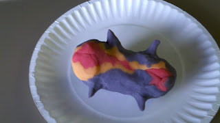
Today in health we learned about how the embryo develops using playdough. Check it out below:
Modeling Embryogenesis
Embryogenesis is the growth and development of the embryo. An important point in embryonic development occurs at the beginning of the third week when the implanted blastocyst differentiates into three spate layers of cells that develop into very specific functions in the human organism.
1. Flatten the three balls of play dough.
2. Holding one disc horizontally (purple), imagine that it is connected by a bubble on top and a bubble on bottom. The top bubble in the amniotic cavity and the bottom is the yolk sac.
a. Amniotic cavity contains amniotic fluid that regulates the temperature of the developing organism and provides a protective cushion from sharp movements
b. The yolk sac produces blood cells until other organs are fully developed and able to take over this function
3. The original disc represents the ectoderm (purple).
a. The ectoderm is a layer of cells that will eventually become the nervous system and the skin.
4. The disc of the inner cell mass differentiates into two layers (place a second flattened ball of red play dough beneath the first – without fusing them). The new layer represents the endoderm (red).
a. The cells in the endoderm are destined to be the digestive system, the lungs, urinary tract, and the glands.
5. A third layer develops between the first 2 (slide the third orange disk between the existing layers of play dough). This final layer represents the mesoderm.
a. The cells in the mesoderm will differentiate into the muscles, the skeleton, the circulatory system, and other internal organs.
6. At the same time that the mesoderm appears, the neural tube begins to form (press the 3 layers together and then form the neural tube down the center of the ectoderm by pinching together two parallel ridges). In the developing embryo, this develops into a hollow tube.
a. The neural tube is the place where the spinal cord and the brain will develop.
7. Next, the edges of the disc begin to fold down around the shrinking yolk sac – resulting in a tube-like/burrito-shaped embryonic body (fold the three layers with the ectoderm (purple)on the outside and the endoderm (red) in the center.
8. Note the three original layers of cells and their locations after this step in embryogenesis.
9. The tube – like body bends into a C-shape (cephalo-caudal folding)
a. Cehpalo – head
b. Caudal – tail